
However, both constructs had the same capacity to produce a glycosaminoglycan (GAG) –specific extracellular matrix.Ĭitation: Adesida AB, Mulet-Sierra A, Laouar L, Jomha NM (2012) Oxygen Tension Is a Determinant of the Matrix-Forming Phenotype of Cultured Human Meniscal Fibrochondrocytes. Furthermore, the constructs under hypoxia produced a matrix with higher mRNA ratio of aggrecan to versican (3.5-fold, p<0.05). This was confirmed by enhanced deposition of collagen II using immuno-histochemistry. The results showed that constructs under normoxia produced a matrix with enhanced mRNA ratio (3.5-fold higher p<0.001) of collagen type II to I. After 3 weeks of in vitro culture, the constructs were assessed biochemically, histologically and for gene expression via real-time reverse transcription-PCR assays. Constructs containing normoxia-expanded MFC were subsequently cultured under normoxia while those formed from hypoxia-expanded MFC were subsequently cultured under hypoxia.
#OPTIXCAM DEFINITION FREE#
The MFC seeded scaffolds (constructs) were cultured in a serum free chondrogenic medium for 3 weeks under normoxia and hypoxia. Normoxia and hypoxia expanded MFC were seeded on to a collagen scaffold.
#OPTIXCAM DEFINITION SOFTWARE#
Check the software settings for "exposure".
#OPTIXCAM DEFINITION DRIVERS#

If it is slightly between positions the image will not be projected up to the camera properly. If you are using a high power compound biological microscope, make sure that your objective lens is clicked all the way into position. However, cameras require extra light in order to be able to pick up that same image, so make sure you have a brightly lit sample under the microscope.Īre objectives clicked into place and caps removes off stereo objectives? Many times when you look through a stereo microscope without the light on you might still be able to see the image. Particularly on a stereo microscope - is the light on?

Many camera sensors are sensitive and if your image is not in focus the camera might not pick it up. The beam splitter will either be a switch or a lever you can pull out and push in.ĭo you have a specimen under the microscope and in focus?īefore you try to view images through the camera or on your computer screen, make sure that your sample is under the microscope, the light is turned on and it is in focus. You will find the beam splitter on the side of the head of your microscope. When the beam splitter is not pulled out or engaged, the light will not travel up to the camera and you will not be able to view an image at the camera.

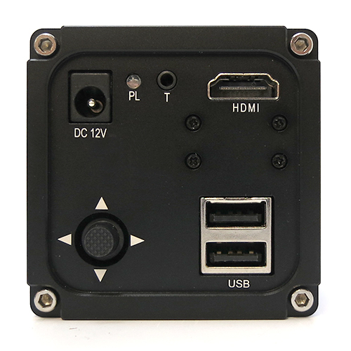
The microscope beam splitter directs light up the trinocular port to the camera. Is your microscope beam splitter engaged? Below you will find some troubleshooting tips for viewing live images on the camera or computer monitor from your digital microscope. However, there is nothing more frustrating than not being able to view an image on your computer monitor from your digital microscope. Digital microscopy and microscope cameras and software are fantastic additions to the world of microscopy.


 0 kommentar(er)
